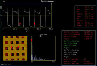
 Section analysis of a standard 10 micron grid. You can see clearly the recesses every 10 microns.
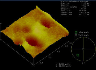
 A 3D image derived from contact mode AFM. This means that the tip and sample are actually "touching" each other.
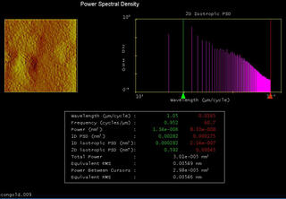
 Power spectral density image of contact gold. This time I'm using a picture derived from the phase difference between some signals. (I also blur) Don't ask me what is PSD, it just gives a lot of information.
  A flattened image of the standard grid. Look at the phase picture on the right. It provides fricking accurate pictures of the surface characteristics. In this case, you can see the roughness on the surface.
Since these experiments were all performed by me, please do not infringe copyright laws. MY VERY FIRST OWN COPYRIGHT PICTURES!!!
No comments:
Post a Comment